
|
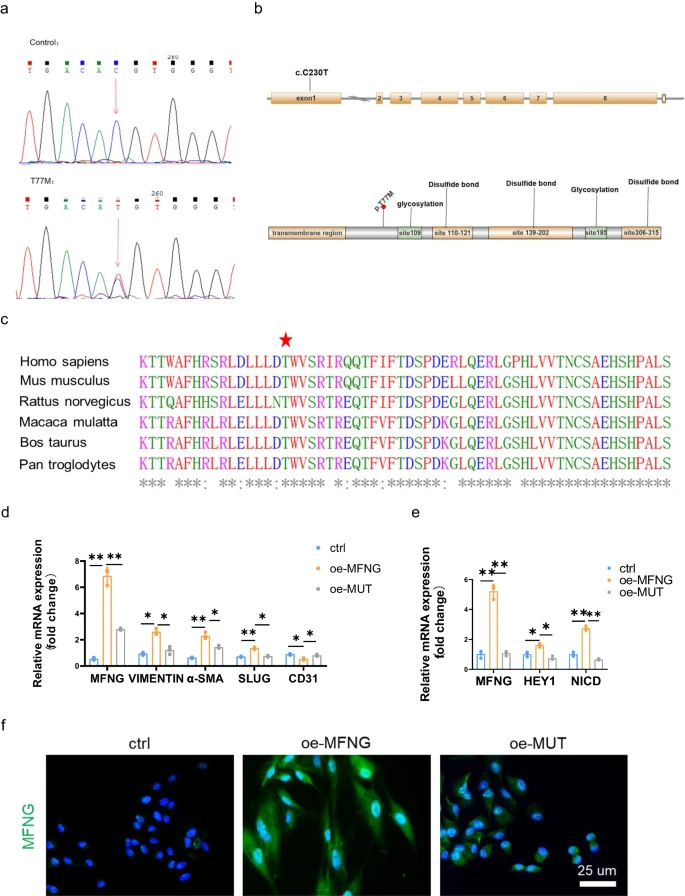
Fig. 7 Validation and in silico analysis of the MFNG variant. a Sequences showing the MFNG missense variant, identified in both patients and healthy controls, with arrows indicating heterozygous nucleotide changes. b Depiction of the MFNG gene’s exon and protein structure, highlighting the location of the genetic variation within this study. Exon 1 of MFNG harbored the base substitutions. The amino acids were primarily composed of glycosylation and disulfide bond domains. c The MFNG mutation was found to exhibit a high degree of conservation across vertebrates, as evidenced by the Clustal X alignment of the MFNG protein in various species. Stars indicate identical residues. In our study, human umbilical vein endothelial cells (HUVECs) served as the control group. HUVECs transfected with the MFNG wild-type plasmid were designated as the overexpression group (oe-MFNG), while HUVECs transfected with the MFNG mutant plasmid were designated as the mutant group (oe-MUT). d, e The relative mRNA expression of MFNG, genes associated with EndMT, and molecules related to the Notch pathway in HUVECs subjected to various treatments was meticulously assessed, employing GAPDH as a reference gene. Significantly distinct mRNA expression levels of MFNG, EndMT-related genes, and Notch pathway molecules were evident when comparing the oe-MFNG and oe-MUT groups. f Cellular immunofluorescence staining underscored the diminished expression of MFNG in the oe-MUT groups compared to the oe-MFNG groups, as visualized through microscopic images. Scale bars = 25 µm