
|
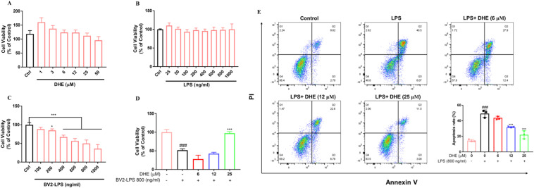
Fig. 5 DHE exhibited neuroprotective effects on SH-SY5Y cells against LPS-stimulated microglia-mediated neurotoxicity. (A, B) SH-SY5Y cells were treated with the indicated DHE (A) or LPS (B) concentrations. (C) SH-SY5Y cells were treated with conditioned medium from BV2 cells that had been exposed to 25–1000 ng/ml of LPS for 24 h, and the cell viability was detected by the MTT assay. (D) SH-SY5Y cells were treated with condition medium from BV2 microglia exposed to LPS (800 ng/ml) for 24 h after pretreatment with DHE (6, 12, 25 μM) for 1 h. The cell viability was detected by the MTT assay. (E) SH-SY5Y cells were co-cultured with BV2 cells in the presence of DHE (6, 12, 25 μM) for 1 h, followed by LPS stimulation for 24 h. Apoptosis was quantified by flow cytometry. Data are presented as the mean ± SD analyzed by one-way ANOVA. ∗p < 0.05 and ∗∗∗p < 0.001 vs. the LPS-treated group, ###p < 0.001 vs. the control group; n = 3.