IMAGE

FIGURE 7

ID
ZDB-IMAGE-250411-8
Source
Figures for Strachan et al., 2025
Image
Figure Caption

FIGURE 7

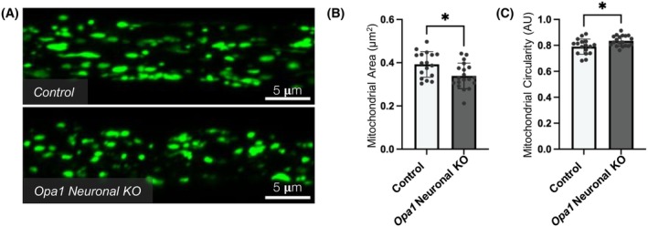
Altered mitochondrial morphology within motor neuron axons in Opa1 neuronal KO Drosophila. (A) Representative confocal images of mitochondria (green) within motor neuron axons. Larvae are progeny of UAS‐Cas9;d42GAL4.UAS‐mitoGFP flies crossed to w1118 (control) or Opa1 sgRNA (Opa1 neuronal KO) flies. Graphs represent mean ± SD mitochondrial size (B) and circularity (C). Statistics consist of two‐tailed t‐tests.

Acknowledgments
This image is the copyrighted work of the attributed author or publisher, and ZFIN has permission only to display this image to its users. Additional permissions should be obtained from the applicable author or publisher of the image. Full text @ FASEB J.