
|
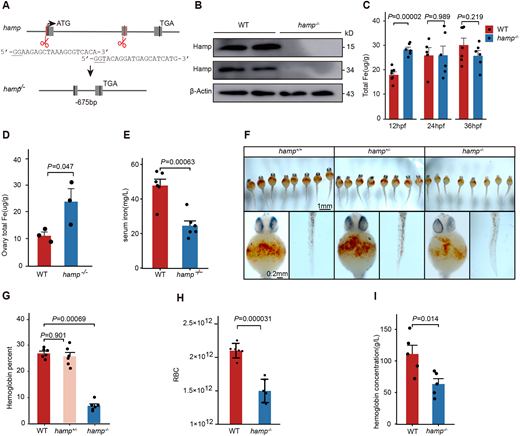
Fig. 1 Deletion of the zebrafish hamp gene reduces hemoglobin synthesis during the embryonic period. (A) Schematic of the targeting site in hamp and the resulting nucleotide sequence in the mutant. (B) Hamp protein level in the wild-type and hamp−/− embryos detected using anti-Hamp antibody. (C) Total iron content of wild-type and hamp−/− embryos at 12 hpf, 24 hpf and 36 hpf (n=6, unpaired, two-tailed Student's t-test). (D) Total iron content of wild-type and hamp−/− ovaries (n=3, unpaired two-tailed Student's t-test). (E) Serum iron of wild-type and hamp−/− peripheral blood (n=6, unpaired, two-tailed Student's t-test). (F) O-Dianisidine staining of functional hemoglobin in the mature primitive erythrocytes in wild type (left), hamp+/− (middle) and hamp−/− (right) at 36 hpf. (G) The hemoglobin levels of embryos was significantly reduced in hamp−/− compared with wild type at 36 hpf (n=6, unpaired, two-tailed Student's t-test). (H) The number of erythrocytes was significantly reduced in hamp−/− compared with wild type at 8 mpf (n=6, unpaired, two-tailed Student's t-test). (I) Hemoglobin concentration was significantly reduced in hamp−/− compared with wild type in adult peripheral blood (n=5, unpaired, two-tailed Student's t-test). Data are mean±s.e.m.