
Image
|
Figure Caption
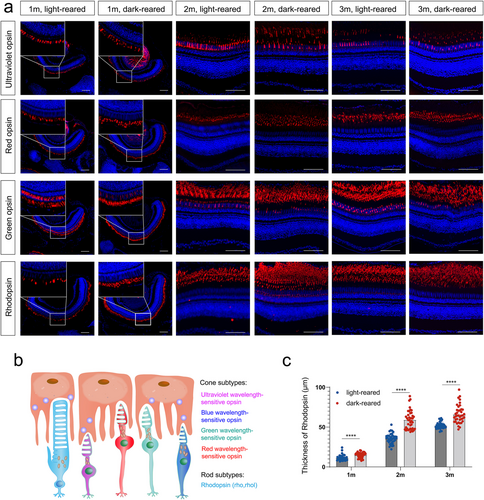
Fig. 3 Expression of opsin in light- and dark-reared zebrafish retinas (a) Immunostaining of retinal paraffin sections, in which opsins are labeled in red and DAPI is marked in blue. The scale bars are 50 μm. (b) The pattern of zebrafish opsin isoforms, including four types of cone opsin and one type of rod opsin. (c) Quantitative measurement of rhodopsin thickness in zebrafish at 1, 2, and 3 months. n = 8 eyes per group at each time point. Statistical comparisons between individual groups were carried out using the Student's t-test. m, month; DAPI, 4′,6-diamidino-2-phenylindole. ****p < 0.0001
Acknowledgments
This image is the copyrighted work of the attributed author or publisher, and
ZFIN has permission only to display this image to its users.
Additional permissions should be obtained from the applicable author or publisher of the image.
Full text @ FASEB J.