
|
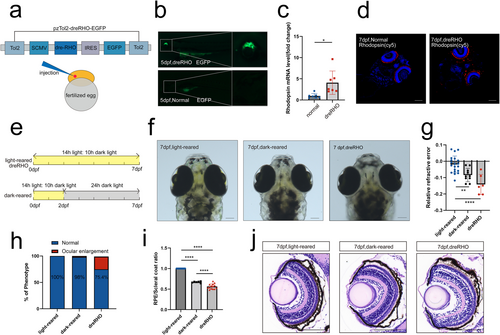
Fig. 4 Rhodopsin overexpression led to RRE changes and scleral enlargement in zebrafish larvae. (a) dreRHO structure. Plasmids are introduced by microinjection into the yolk sacs of fertilized eggs. (b) EGFP expression is observed in zebrafish at 5 dpf. (c) Rhodopsin mRNA levels of zebrafish at 7 dpf. n = 7 eyes per group. (d) Rhodopsin immunofluorescence staining (4D2). (e) Light cycles in three groups of zebrafish. (f) Zebrafish photographed under a microscope at 7 dpf. (g) Relative refractive error at 7 dpf; n = 20, 10, and 10 eyes for light-reared, dark-reared, and dreRHO group, respectively. (h) Proportion of ocular enlargement phenotypes. n = 600, 600, and 200 eyes for light-reared, dark-reared, and dreRHO group, respectively. (i) Ratio of RPE diameter to scleral diameter; n = 20, 6, and 14 eyes for light-reared, dark-reared, and dreRHO group, respectively. (j) Results of HE staining on the eyeballs at 7 dpf. Statistical significance was determined using the Student's t-test and the one-way ANOVA with Bonferroni correction (more than two groups). The scale bars are 100 μm. ANOVA, analysis of variance; dreRHO, rhodopsin expression plasmid; EGFP, enhanced green fluorescent protein; HE, hematoxylin–eosin; mRNA, messenger ribonucleic acid; RPE, retinal pigment epithelial. *p < 0.05, **p < 0.01, ****p < 0.0001.