
|
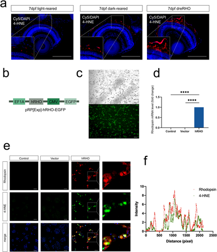
Fig. 6 Rhodopsin induces accumulation of 4-HNE in the retinal pigment epithelium (a) Immunostaining of eye paraffin sections in which 4-HNE is labeled in red and DAPI is marked in blue. (b) Plasmid structure of rhodopsin. (c) Plasmid EGFP fluorescence and rhodopsin expression levels after transfection. (d) Rhodopsin mRNA levels detected using qRT-PCR. (e) Immunofluorescence. (f) Fluorescence colocalization analysis. Scale bars are 50 μm. 4-HNE, 4-hydroxynonenal; DAPI, 4′,6-diamidino-2-phenylindole; EGFP, enhanced green fluorescent protein; mRNA, messenger ribonucleic acid; qRT-PCR, quantitative reverse transcriptase polymerase chain reaction. ****p < 0.0001.