
|
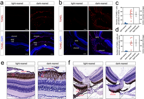
Fig. 8 TUNEL and HE staining of 3-month-old zebrafish (a, b) TUNEL staining of the peripheral retina (n = 9 for the light-reared group and n = 12 for the dark-reared group) and optic nerve (n = 7 for the light-reared group and n = 9 for the dark-reared group). The white arrow indicates the migration of the RPE with blue fluorescence marked by DAPI. (c, d) Statistical results of TUNEL staining. (e, f) HE staining suggested thinning of the choroid and RPE migration (shown by the red arrow). The scale bars are 100 μm. DAPI, 4′,6-diamidino-2-phenylindole; HE, hematoxylin–eosin; RPE, retinal pigment epithelium; TUNEL, terminal deoxynucleotidyl transferase-mediated dutp nick-end labeling.