IMAGE

Figure 6

ID
ZDB-IMAGE-250728-24
Source
Figures for Boi et al., 2025
Image
Figure Caption

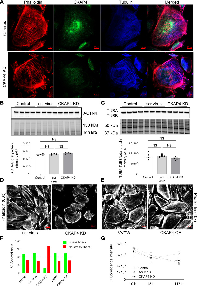
Figure 6 CKAP4 KD in HPODs in vitro alters the cytoskeleton.

Immunofluorescence staining of HPODs with phalloidin (red, actin fibers), tubulin (blue, microtubules), and CKAP4 (green) in scr control and CKAP4 KD. HPODs showed loss of actin stress fibers and rearranged microtubules (A). ACTN4 (B) and TUBA-TUBB (C) protein expressions were investigated by Western blot. Total protein blots and normalized protein expression graphs are also presented. Representative pictures of phalloidin staining of scr control and CKAP4 KD HPODs (D) and VVPW control and CKAP4 OE (E). Percentage of cells scored for the presence or absence of stress fibers in both CKAP4 KD and -OE experiments (F). HPOD viability in untreated, scr control, and CKAP4 KD cells from transfection time to 117 hours (G). F: a minimum of 64 cells per group were scored, and percentages are reported in the graph. G: n = 8 per time point/group, Friedman’s test was used, NS. AU, arbitrary units; CKAP4, cytoskeleton associated protein 4; KD, knockdown; OE, overexpression; HPODs, human podocytes; scr, scrambled; ACTN4, αactinin-4; TUBA, tubulin-α; TUBB, tubulin-β.

Acknowledgments
This image is the copyrighted work of the attributed author or publisher, and ZFIN has permission only to display this image to its users. Additional permissions should be obtained from the applicable author or publisher of the image. Full text @ JCI Insight