
|
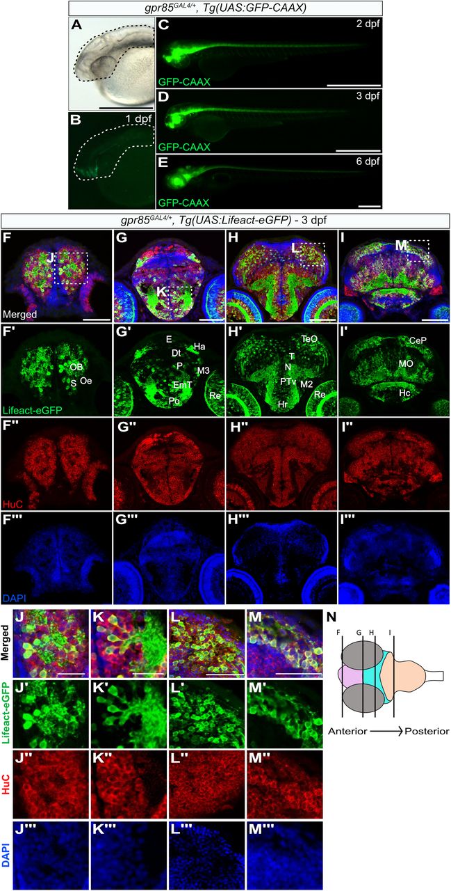
Fig. 1 Gpr85 is broadly expressed by maturing neurons in the zebrafish brain parenchyma during late embryogenesis. A–E, Lateral views of live gpr85GAL4/+, Tg(UAS:GFP-CAAX) zebrafish embryos at 1 dpf with bright-field (A) and fluorescence imaging (B–E) showing the GFP-CAAX signal in the brain and spinal cord at 1 dpf (B), 2 dpf (C), 3 dpf (D), and 6 dpf (E). (N = 3) Scale bars, 50 μm. F–I, Maximum projection confocal images of coronal sections of the brain from a 3 dpf gpr85GAL4/+, Tg(UAS:Lifeact-eGFP) zebrafish larva stained with anti-GFP (green; F′–I′), anti-HuC (red; (F″–I″) and DAPI (blue; F″′–I″′). Lifeact-eGFP+ cells are observed in the OB, subpallium (F′), Ha, retina, eminentia thalami, preoptic region, pallium (G′), tectum opticum, hypothalamus (H′), cerebellar plate, and MO (I′). Boxed regions are enlarged in panels J to M. Scale bars, 50 µm. J–M, Enlarged optical sections of the areas boxed in panels (F–I), showing Lifeact-eGFP+/HuC+ neurons in the brain parenchyma. (N = 3) Scale bars, 25 µm. N, Schematic representation of the coronal sections of the brain illustrated in F–I, with the forebrain in pink, the midbrain in turquoise, and the hindbrain in peach. CeP, cerebellar plate; DT, dorsal thalamus; EmT, eminentia thalami; H, rostral hypothalamus; Ha, habenula; Hc, caudal hypothalamus; MO, medulla oblongata; N, region of the nucleus of medial longitudinal fascicle; M2, migrated posterior tubercular area; M3, migrated area of EmT; OB, olfactory bulb; Oe, olfactory epithelium; P, pallium, Po, preoptic region; PTv, ventral part of posterior tuberculum; Re, retina; S, subpallium; TeO, tectum opticum; T, midbrain tegmentum; dpf, days postfertilization.