
|
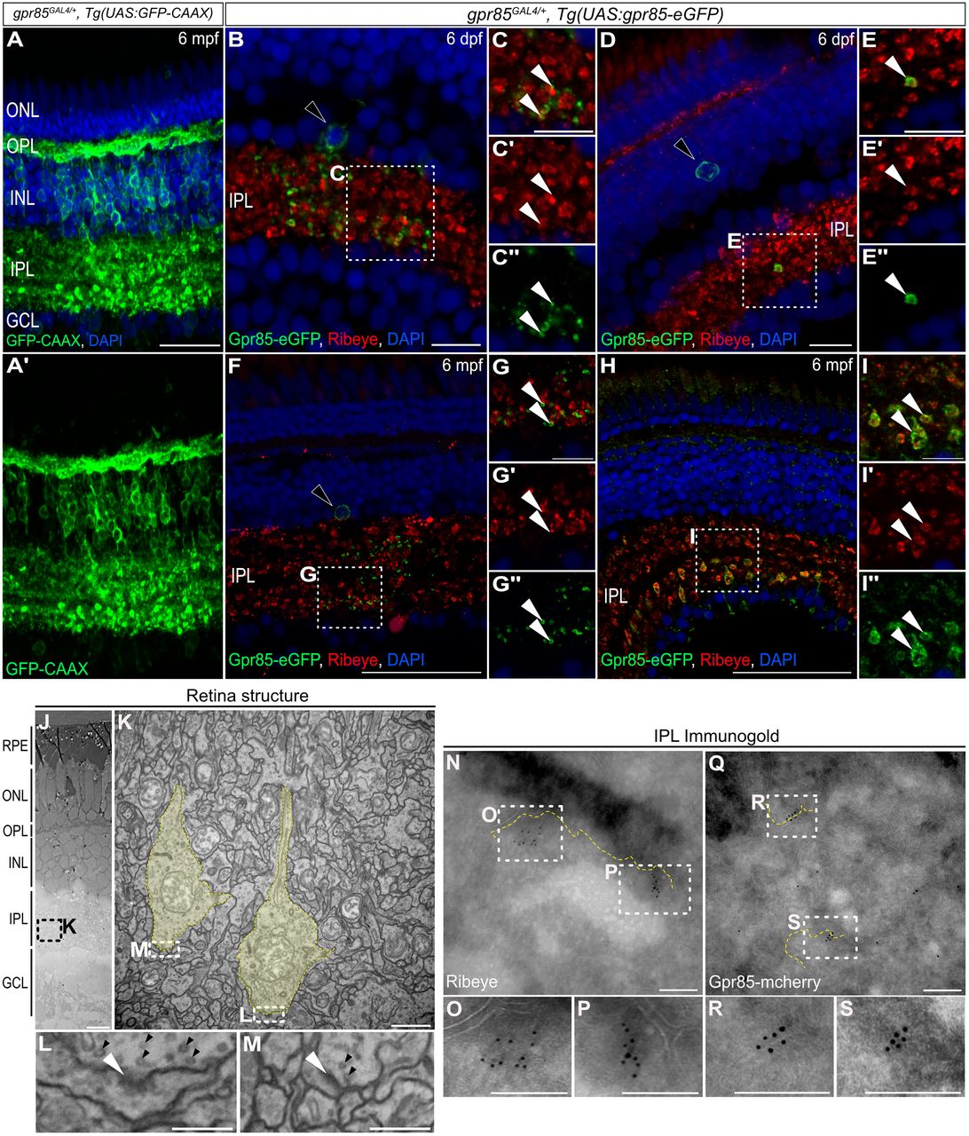
Fig. 3 Gpr85 is enriched in the pre- and postsynaptic compartments of developing and adult IPL retinal ribbon synapses. A, Maximum projection confocal images of the retina from a gpr85GAL4/+, Tg(UAS:GFP-CAAX) adult zebrafish stained with anti-GFP (green) and DAPI (blue). (N = 3) Scale bar, 50 µm. B–I, Confocal images of coronal retinal sections from 6 dpf (B–E″) or 6 mpf (F–I″) gpr85GAL4/+, Tg(UAS:gpr85-eGFP) zebrafish stained with anti-GFP (green), anti-Ribeye-A (red), and DAPI (blue). B, F, Larval or adult Gpr85-eGFP+ AC somas and signal within the IPL are shown, with boxed regions enlarged in C-C″ and G-G″, respectively. White arrowheads highlight Gpr85-eGFP signals adjacent to Ribeye+ ribbon terminals. D, H, Larval or adult Gpr85-eGFP+/Ribeye-A+ BPC ribbon presynaptic terminals are shown, with boxed regions enlarged in E-E″ and I-I″, respectively. White arrowheads show examples of Gpr85-eGFP signal present in Ribeye+ ribbon terminals. (N = 3) Scale bars, (B, D) 25 µm, (F, H) 50 µm, (C, E, G, I) 10 µm. J, Electron microscopy coronal view of the structure of 6 dpf retina from gpr85GAL4/+, Tg(UAS:gpr85-mCherry) zebrafish larvae. (N = 3) Scale bar, 10 µm. K, Enlarged image of the ribbon presynaptic terminals (yellow) from the boxed area in panel J. Scale bar, 1 µm. L, M, Enlarged images of synaptic boutons from the boxed area in panel K. White arrowheads highlight postsynaptic densities while black arrowheads point to presynaptic vesicles. Scale bars, 200 nm. N–S, Immunogold labeling of Ribeye (N–P) and mCherry-tagged Gpr85 (Q–S) focusing on the retinal inner plexiform layer from 6 dpf gpr85GAL4/+, Tg(UAS:gpr85-mCherry) larvae. Dashed yellow lines highlight plasma membranes. Boxes indicate regions enlarged in panels O, P, R, and S. Scale bars, 200 nm. dpf, days postfertilization; mpf, months postfertilization; ONL, outer nuclear layer; OPL, outer plexiform layer; INL, inner nuclear layer; GCL ganglion cell layer.