
|
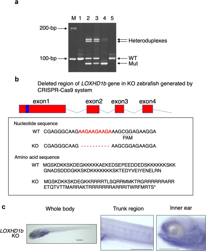
Fig. 2 (a) Heteroduplex mobility assay (HMA). Lane 5 indicates WT (control). In lane 1, the same band as in lane 5 appeared. Lane 2 and lane 3 indicate heterozygous mutants. Lane 4 indicates a band shorter than lane 1 (WT), representing a homozygous mutant. (b) Mutated LOXHD1b loci in KO zebrafish generated using the CRISPR-Cas9 system. gRNA was created in the region adjacent to the PAM sequence in the first exon. Sequencing of the homozygous mutant revealed deletion of 10 bases in the first exon. Moreover, translation of nucleotide sequence to amino acid sequence after the deletion of 10 nucleotides revealed the formation of a premature stop codon. (c) LOXHD1b mRNA expression in KO zebrafish assessed using in situ hybridization. The spine and periocular organs were labeled with an antisense RNA probe in KO zebrafish. Scale bar = 300 μm for C