
Image
|
Figure Caption
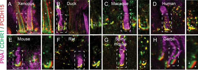
Figure 2
Evolutionarily conserved localization of pcdh15 and cdhr1 in photoreceptors predicts interactions linking outer segments and calyceal processes.
Structured illumination microscopy (SIM) of wildtype xenopus (A), mallard duck (B), macaque (C), human (D), mouse (E), rat (F), spiny mouse (G), and gerbil (H), retinal sections probed for cdhr1 (green), pcdh15 (red), and PNA (magenta) to label cone outer segments. White boxes represent the inset enlargements. White arrowheads highlight the juxtaposition of the cdhr1a and pcdh15b signals in each species. Scale bar = 5 μm.
Acknowledgments
This image is the copyrighted work of the attributed author or publisher, and
ZFIN has permission only to display this image to its users.
Additional permissions should be obtained from the applicable author or publisher of the image.
Full text @ Elife