
FIGURE
Figure 2
- ID
- ZDB-FIG-260418-14
- Publication
- Patel et al., 2026 - Cdhr1a and pcdh15b may link photoreceptor outer segments with calyceal processes revealing a potential mechanism for cone-rod dystrophy
- Other Figures
- All Figure Page
- Back to All Figure Page
Figure 2
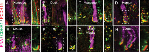
Evolutionarily conserved localization of pcdh15 and cdhr1 in photoreceptors predicts interactions linking outer segments and calyceal processes. |
Expression Data
Expression Detail
Antibody Labeling
Phenotype Data
Phenotype Detail
Acknowledgments
This image is the copyrighted work of the attributed author or publisher, and
ZFIN has permission only to display this image to its users.
Additional permissions should be obtained from the applicable author or publisher of the image.
Full text @ Elife