FIGURE SUMMARY
Title

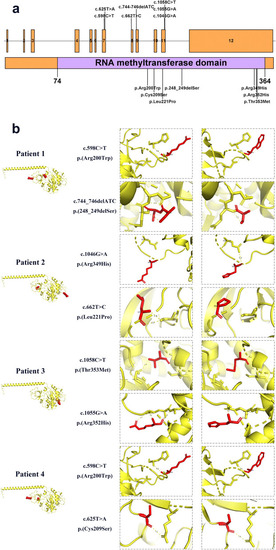
SPOUT1 variants associated with autosomal-recessive developmental and epileptic encephalopathy

Authors
Liu, W., Gao, K., Du, X., Wen, S., Yan, H., Wang, J., Wang, Y., Song, C., Lin, L., Ji, T., Gu, W., Jiang, Y.
Source
Full text @ Acta Epileptol

SPOUT1 gene, protein regions and variant sites. b Protein structures of different SPOUT1 variants as predicted by AlphaFold

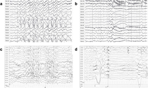
Electroencephalography (EEG) of patients with SPOUT1 variant. Interictal EEG and ictal scalps of Patient 2 (a, b) and Patient 4 (c, d) demonstrating atypical hypsarrhythmia (a, c) and monitored epileptic spasms (b, d)

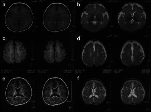
Brain MRI of Patient 1 at age of 5 months (a, b), 1 year and 3 months (c, d) and 2 years and 11 months (e, f). Agenesis of corpus callosum, bilateral frontal atrophy, and white matter hypomyelination were shown

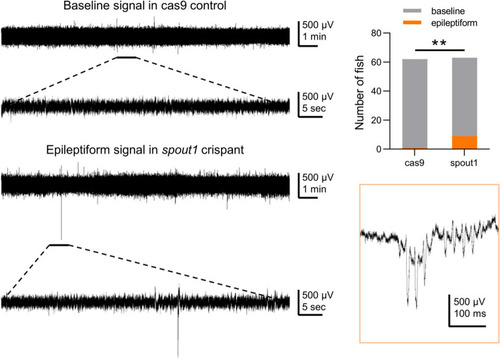
Representative electrophysiological recordings and statistical analysis of zebrafish between the cas9-control group and spout1-knockout group. **P < 0.01

RNAseq analysis of spout1-knockout zebrafish. a Volcano map of gene expression changes after spout1 knockout. Red dots represent up-regulated genes. Green dots represent downregulated genes. b Significantly enriched GO terms, which were grouped into three categories: molecular function (MF), cellular component (CC) and biological process (BP). c GOCircle plot based on the GOplot analysis of enriched GO term. The inner ring is a barplot where the height of the bar indicates the significance of the GO term (-log10 |P-value|), and color corresponds to the z-score, which indicates increase (red) or decrease (blue). The outer ring displays scatterplots of the expression levels (logFC) of the genes in each term

ZFIN is incorporating published figure images and captions as part of an ongoing project. Figures from some publications have not yet been curated, or are not available for display because of copyright restrictions.

Acknowledgments
This image is the copyrighted work of the attributed author or publisher, and ZFIN has permission only to display this image to its users. Additional permissions should be obtained from the applicable author or publisher of the image. Full text @ Acta Epileptol