
Figure 2
- ID
- ZDB-FIG-230615-7
- Publication
- Schellens et al., 2023 - A protein domain-oriented approach to expand the opportunities of therapeutic exon skipping for USH2A-associated retinitis pigmentosa
- Other Figures
- All Figure Page
- Back to All Figure Page
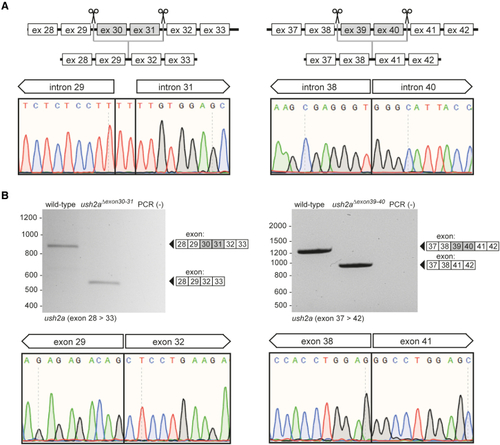
Design and characterization of the ush2aΔexon30-31 and ush2aΔexon39-40 zebrafish line (A) Schematic representation of the exon-excision approach. Sanger sequencing confirmed the presence of the anticipated excisions in injected embryos (1 day post fertilization [dpf]). Excision of the genomic region containing ush2a exons 30 and 31 resulted in the insertion of two nucleotides (TT) at the repair junction. (B) RT-PCR analysis revealed the absence of ush2a exons 30 and 31 in ush2aΔexon30-31 larvae and the absence of exons 39 and 40 in ush2aΔexon39-40 larvae (5 dpf). Sanger sequencing of the ush2aΔexon30-31 and ush2aΔexon39-40 amplicons confirmed the absence of the target exons from the transcript. |