|
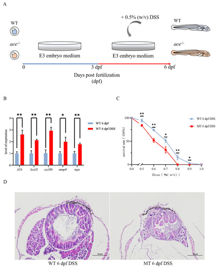
The IBD model was successfully constructed using DSS. (A) A diagram of IBD model development. (B) Changes in DSS-treated and untreated wild-type larvae were examined by qRT-PCR for several pro-inflammatory factors: ilb, lect2l, cxcl8b, mmp9, and mpx. Data represent the mean ± SD. * p < 0.05, ** p < 0.01. Three independent biological replicates were performed. (C) Line chart of survival rates comparing ace−/− mutants and wild-type larvae treated as the DSS dose (% (w/v)) increased from 0% to 1% (n = 50). The significance of differences is annotated above the nodes in the line chart. * p < 0.05, ** p < 0.01. (D) HE staining shows a more severely disorganized intestinal epithelium in ace−/− mutants compared with the wild type at 6 dpf following DSS treatment.
|