
Fig. 2
- ID
- ZDB-FIG-240703-2
- Publication
- Webb et al., 2021 - EHD2 modulates Dll4 endocytosis during blood vessel development
- Other Figures
- All Figure Page
- Back to All Figure Page
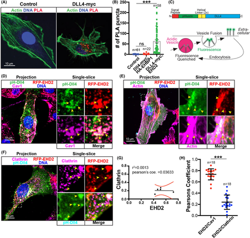
Membranous Dll4 localizes with EHD2 and caveolin-1. (A) Representative image of proximity ligation assay (PLA). Cells were stained as indicated and PLA reaction is marked by red puncta. Control condition was not transfected. Dll4-myc condition was only transfected with Dll4-myc and stained for endogenous EHD2. (B) Graph of number of PLA puncta by condition. Control condition was not transfected. EHBP1 condition was transfected with EHBP1-HA and Dll4-myc. Dll4-myc condition was only transfected with Dll4-myc and stained for endogenous EHD2. (C) Schematic of engineered Dll4 (top). Bottom, cartoon of pH-dependent function of GFP variant pHluorin tag. PHluorin fluoresces on the membrane at neutral pH but is quenched when internalized into acidic endosomes allowing for visualization of only membranous Dll4. (D) Representative image of endothelial cell (HUVEC) stained for caveolin-1 (Cav1) expressing pHluorin-Dll4 (pH-Dll4) and RFP-EHD2. (E) Representative image of cell stained for actin expressing pH-Dll4 and RFP-EHD2. (F) Representative image of cell stained for clathrin expressing pH-Dll4 and RFP-EHD2. (G) Proportion of coincidence of clathrin (y-axis) and EHD2 (x-axis) around Dll4 puncta. (H) Pearson's correlation between indicated proteins. N, number of cells. Boxes denote magnified images on right. Yellow arrowheads show areas of pH-Dll4 puncta. ***p < .001. Error bars are 95% confidence intervals. All experiments were done at minimum in triplicate |