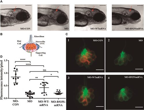
In vivo rescue assay of myo6b MO phenotype with human mRNAs. Coinjection of human wild-type MYO6 mRNA with myo6b MO resulted in partial rescue of the knockdown phenotype, whereas co injection of MO and human MYO6 mRNA with the c.2775G>C p.Arg925Ser novel variant (MO-p.R925S-mRNA) was unable to rescue the knockdown phenotype. (A) Lateral views of the head of 5dpf larvae labeled with 4-Di-2-Asp (10X magnification). Neuromasts stained are shown as white dots. Red arrows point at neuromasts situated between the otic vesicle and the eye, where fluorescence intensity measurements were performed. Scale bar: 200 μm. (B) Schematic depicts a lateral view of a neuromast. (C) Confocal images showing the uptake of 4-Di-2-Asp (red) by LL hair cells of 5 dpf Tg (Brn3c:GFP) larvae. GFP (green) is expressed in neuromast hair cells. Scale bar: 9 μm. (D) Box and whisker plot: fluorescence intensity per pixel (F/pix) normalized to the fluorescence intensity of non-injected larvae. ANOVA followed by Tukey multiple comparison-test,: ****P < 0.0001, ***P< 0.001, **P< 0.01, *P < 0.05, n = 10 neuromasts from five larvae, for each group.
|
