|
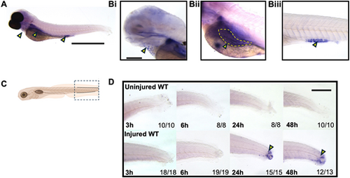
Wholemount in situ hybridization demonstrates ctsz expression in cells in the injured tailfin. (A-Biii) In situ hybridization of ctsz in unchallenged larvae at 3 dpf (A) highlighting cells overlying the heart cavity (Bi), the intestinal bulb (Bii) and mid-intestine (Biii). (C) Tailfin injury schematic view demonstrating the analysed area in the in situ hybridization of ctsz experiments. (D) Representative images of in situ hybridization of ctsz in unstimulated wild-type (WT) (top row) and injured WT (bottom row) larvae at different time points. Numbers at bottom right indicate the number of analysed animals. Yellow arrowheads indicate staining at recruited individual leukocytes. Yellow dashed line outlines the intestinal bulb. Scale bars: 1 mm (A); 500 µm (D); 330 µm (Bi).
|