
Figure 1
- ID
- ZDB-FIG-260623-10
- Publication
- Capecki et al., 2026 - Evidence that disruption of Discoidin domain receptor 2 contributes to palate malformations through effects on the extracellular matrix
- Other Figures
- All Figure Page
- Back to All Figure Page
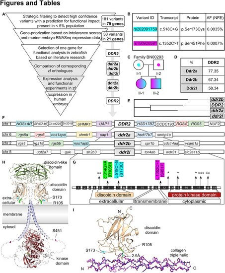
Selection of a suitable nsCPO candidate gene for analysis in zebrafish models. (A) workflow from exome sequencing (ES) reanalysis of data from nsCPO sib-pairs (white) to functional experiments in the zebrafish model (grey). (B) DDR2 variants in our index family (BN00293). Each parent carried a different variant in DDR2. Allele frequencies (AF) were obtained from GnomAD v.4.1.0. (C) index family pedigree. The parents were unaffected, while both children have nsCPO and are compound-heterozygotes for the parental variants. (D–F) comparative analysis of human DDR2 and zebrafish ddr2a, ddr2b, and ddr2l. Comparison of alignment of Ddr2a, Ddr2b, and Ddr2l with the human DDR2 amino acid sequence (D) and phylogenetic relationships (E). Comparison of human DDR2 and zebrafish ddr2a, ddr2b, and ddr2l genetic loci: Corresponding genes are presented in the same color (F). (G) human DDR2 gene structure: Exons are shown approximately to scale, relative to the entire gene. Protein domains are marked according to coding exons. Previously reported variants are indicated with arrows in their corresponding exons: * and °, variants associated with SMED and Warburg-Cinotti syndrome, respectively. The two variants identified in our study are shown (c.518C>G, c.1352C>T), as well as one further variant potentially causing a cleft palate from the DECIPHER database (c.314G>A). For further information refer to the text and the appendix (SI Appendix, Table S2). (H) localization of three potential causative variants in the DDR2 protein. AlphaFold3 model of a human DDR2 dimer, based on individual modeling of the N-terminal discoidin (29–185), discoidin-like (190–370), transmembrane helix (400–420), and C-terminal tyrosine-kinase (563–849) domains. Positions of the three variant sites are labeled (R105, S173, S451). (I) variants R105H and S173C in the discoidin domain are in the collagen triple-helix binding interface. R105 forms a hydrogen-bond with a hydroxyproline, which cannot be substituted by the shorter histidine sidechain. S173 is close to two prolines of the collagen-helix and to a disulfide bond (C177–C73), which stabilizes the extracellular domain. Hydrogen residues are red, nitrogen blue. N, N-term; C, C-term. |