IMAGE

FIGURE 2

ID
ZDB-IMAGE-250306-20
Genes
Source
Figures for Liu et al., 2024
Image
Figure Caption

FIGURE 2

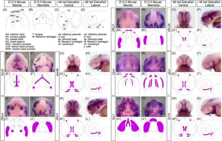
Comparing the expression pattern of the selected genes during craniofacial morphogenesis in mouse and zebrafish embryos. (A–D) Schematic illustrations of the craniofacial anatomy of the maxillary (A) and mandibular (B) regions of mice at E12.5 and zebrafish at 48 hours postfertilization (hpf) (C,D). (E–X) The expression pattern of Sox9 (sox9b), Zfhx4 (zfhx4), Zfhx3 (zfhx3b), Cjun (cjun), and Six1 (six1b) were examined using whole‐mount in situ hybridization. The samples are shown at the top, and the genes are shown on the left. (E′–X′) Schematic summary of the expression pattern of the selected genes. Black arrows, black arrowheads, and white arrowheads indicate Meckel's cartilages, ethmoid plate, and ceratohyal, respectively. Scale bars: 500 μm in V; 200 μm in X.

Figure Data
Acknowledgments
This image is the copyrighted work of the attributed author or publisher, and ZFIN has permission only to display this image to its users. Additional permissions should be obtained from the applicable author or publisher of the image. Full text @ Dev. Dyn.