IMAGE

FIGURE 4

ID
ZDB-IMAGE-250609-57
Antibodies
Source
Figures for Mesika et al., 2025
Image
Figure Caption

FIGURE 4

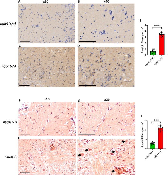
Amyloid aggregation in adult NGLY1‐deficient zebrafish brains. Representative histological images of ngly1(+/+)(A, B, F, G) and ngly1(−/−)(C, D, H, I) zebrafish brain and optic tectum (TeO) reacted with an antibody against amyloid fibrils (AD) and Congo Red (FI), demonstrating significant aggregation of amyloid fibrils (black arrows) at the intracellular space of the mutant fish brain. Magnifications: (F, H): X10, (A, C, G, H): X20, and (B, D): X40. Scale bar: 100 μm. (E, J) Quantification of amyloid fibers by Immunohistochemistry and Congo Red staining, respectively. ***T‐test, p < 0.001 (N = 10 per group).

Figure Data
Acknowledgments
This image is the copyrighted work of the attributed author or publisher, and ZFIN has permission only to display this image to its users. Additional permissions should be obtained from the applicable author or publisher of the image. Full text @ J. Inherit. Metab. Dis.