
|
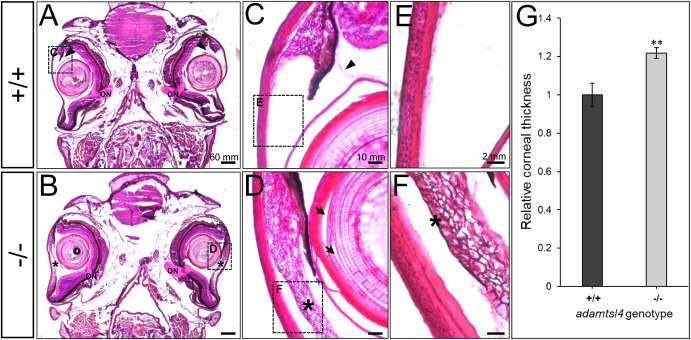
Fig. 7 Histological analysis of hematoxylin-eosin-stained tissue sections from adult adamtsl4 KO zebrafish (5 months). Transverse tissue sections were prepared as described in the Materials and Methods section. Representative photographs of wild type (+/+) and KO (−/−) zebrafish are shown. A-B. Tissue sections from the central part of the eyeball highlighting the presence of the optic nerve (ON). C-D. Anterior segment. E-F. Corneal structure. G. Quantitative analysis of corneal thickness. ∗∗: p < 0.01. Rectangles indicate areas magnified in subsequent panels. Arrows: separation of lens fibers; Asterisks: abnormal annular ligament.
Reprinted from Experimental Eye Research, , Tevar, A., Aroca-Aguilar, J.D., Atiénzar-Aroca, R., Ramírez, A.I., Fernández-Albarral, J.A., Escribano, J., Zebrafish adamtsl4 knockout recapitulates key features of human ADAMTSL4-related diseases: a gene involved in extracellular matrix organization, cell junctions and development, 110572110572, Copyright (2025) with permission from Elsevier. Full text @ Exp. Eye. Res.