
- Title
-
Knockdown of best1 Gene in Zebrafish Caused Abnormal Neuronal and Skeletal Development -A Subtype of Craniovertebral Junction Malformation
- Authors
- Liu, Z., Li, K., Wang, K., Zhang, L., Jia, S., Wang, H., Jian, F., Wu, H.
- Source
- Full text @ Neurospine
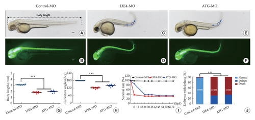
Body length, body axis and survival rate of best1 zebrafish morphants. (A–F) Gross morphology of transgenic (HuC:EGFP) zebrafish embryos at 2-dpf. Compared with control morpholino (MO), knock down best1 causes shorter body length (C–F), curved body axis (C–F). (G) A time-course plot of percent survival in control versus best1 morphants for 3 days. (H) The percentage of embryos with development defects. (I, J) Quantification of body length (I) and curvature angles (J) of embryos. Columns, mean; standard error of the mean (n = 10; analysis of variance), ***p < 0.0001. Scale bar, 100 μm. dpf, days post fertilization. EXPRESSION / LABELING:
PHENOTYPE:
|
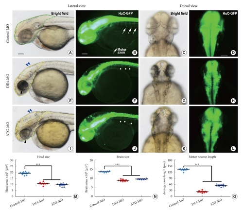
Nervous system developmental abnormalities caused by best1 knockdown. (A–L) Gross morphology of transgenic (HuC:EGFP) zebrafish embryos at 2-dpf. Compared with control morpholino (MO), knockdown of best1 gene causes head and brain patterning defects (E, I, blue arrowheads), microcephaly (E, I, yellow dotted line), small eyes (E, I, black arrowheads), brain-size reductions (F, J, H, and L), and abnormal neuronal outgrowth (F, J, asterisks). (M–O) Quantification of the head area (M), brain area (N) and average motor axon length (O) shows significantly decrease in best1 morphants. Columns, mean; standard error of the mean (n=10; analysis of variance), ***p < 0.0001. Scale bar, 100 μm. dpf, days post fertilization. |
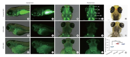
Loss of best1 causes abnormal craniofacial structure and interorbital distance. (A–N) Representative bright field and fluorescent images of zebrafish head skeleton at 6-dpf. In vivo visualization of the skeleton is achieved by the administration of a fluorescent dye (Calcein) directly to the fish water. Dyes that bind to calcified matrix can be used to label the entire skeleton. Lateral view (A–F) and ventral view (G–L) of the head skeleton of day-6 embryos labeled with Calcein. (B, C, E, F, H, I, K, and L) When embryos were injected with best1-morpholino (MO) at the one-cell stage, the amount of stained mineralized tissue was significantly reduced compared to fish injected with control-MO. Panel N show increased intraocular distance (yellow arrows) in best1 morphants. Panel M and N show measurements of the distance between the eyes, and panel O shows the distances depicted graphically as the mean for 10 embryos of each type. Columns, mean; standard error of the mean (n = 10; analysis of variance), ***p < 0.0001. Scale bar, 100 μm. 5ba, fifth branchial arch; op, opercular bone; ec, ectopterygoid; e, ethmoid plate; pq, palatoquadrate; m, Meckel cartilage; dpf, days postfertilization. PHENOTYPE:
|
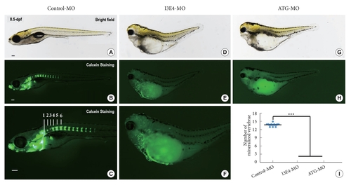
Loss of best1 causes abnormal craniofacial structure and vertebral development. (A–H) Representative bright field and fluorescent images of zebrafish skeleton at 8.5-dpf. In vivo visualization of the skeleton is achieved by the administration of a fluorescent dye (Calcein) directly to the fish water. Dyes that bind to calcified matrix can be used to label the entire skeleton. Lateral view (B, C) of the skeleton and mineralized vertebrae of day-8.5 embryos labeled with Calcein. Vertebrae 1, 2, 3, 4, 5, and 6 are indicated. (E, F, and H) When embryos injected with best1-morpholino (MO) at one-cell stage, the amount of stained mineralized tissue is markedly reduced compared to control-MO-injected fish. Vertebral development is significantly delayed in best1 morphants. (I) Quantification of the number of mineralized vertebrae at 8.5-dpf. Columns, mean; standard error of the mean (n = 10; analysis of variance), ***p < 0.0001. Scale bar, 100 μm. dpf, days postfertilization. PHENOTYPE:
|
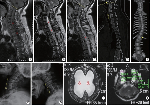
phenotypes of the patient with a premature stop codon of BEST1 gene (p.S79Ffs*153). (A–C) Sagittal magnetic resonance imaging (MRI) revealed Chiari malformation (cerebellar tonsil exceeding the foramen magnum, yellow line) and extensive syringomyelia (red stars) at cervical and thoracic segments. (D–G) Computed tomography and x-ray showed assimilation of atlas (arrow), basilar invagination (odontoid process above the chamberlian line, yellow dotted line), Klippel-Feil malformation (fusion of C5 and C6 vertebrae), butterfly vertebra at T12 (arrow head) and scoliosis. (H, I) MRI of the head indicated hydrocephalus (&) and microphthalmia (diameter of eyeball is smaller than 20 mm). |