
Figure 4
- ID
- ZDB-FIG-210310-116
- Publication
- Harding et al., 2021 - EPHA2 Segregates with Microphthalmia and Congenital Cataracts in Two Unrelated Families
- Other Figures
- All Figure Page
- Back to All Figure Page
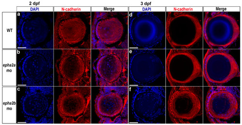
Lens abnormalities in epha2b morphant zebrafish. To assess lens development, N-cadherin (red) and DAPI (blue) staining were performed on lens sections from wild-type (WT), epha2a, and epha2b morphant zebrafish at 2 dpf (a–c) and 3 dpf (d–f); this permitted visualisation of the lens fibre cells and cell nuclei, respectively. At 2 dpf, there were no apparent differences between the morphants and uninjected wild-type siblings. At 3 dpf, the epha2b morphant showed retention of fibre cell nuclei within the lens (f), which was not observed in age-matched wild-type (d) and epha2a morphant larvae. Scale bars = 25 µm. |
| Antibody: | |
|---|---|
| Fish: | |
| Knockdown Reagents: | |
| Anatomical Term: | |
| Stage Range: | Long-pec to Protruding-mouth |
| Fish: | |
|---|---|
| Knockdown Reagents: | |
| Observed In: | |
| Stage Range: | Long-pec to Protruding-mouth |