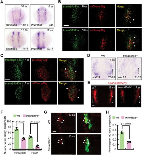
Tmem88 deletion severely impedes PPP specification. (A) In situ hybridization of tmem88a and tmem88b in wild-type embryos. (B, C) Double-color fluorescence in situ hybridization showing the expression of mCherry (red) and tmem88a or tmem88b (green) in Tg(nkx2.3:mCherry) embryos at the 10 (B) and 17 ss (C). The white arrow heads indicate the mCherry+ PPPs that do not express tmem88a or tmem88b. Scale bar, 50 μm. (D) Expression of nkx2.3 in wild-type and tmem88a/b−/− embryos at the 17 ss. The black dotted lines indicate the region where the pouch progenitors are located. (E, F) Representative confocal sections showing mCherry+ PPPs in wild-type and tmem88a/b−/− embryos at 17 ss (E). The PPPs are indicated by white dotted lines. Scale bar, 50 μm. Quantification of the number of pericardial and pouch progenitors positive for mCherry in wild-type and tmem88a/b−/− mutants is shown in (F). The group values are expressed as mean ± SD. (G, H) Requirement of Tmem88 for pouch progenitor specification. Wild-type and tmem88a/b−/− mutant embryos in the Tg(nkx2.3:mCherry;sox17:GFP) background were imaged at 10 ss (G). Scale bar, 20 μm. Ratio of mCherry+ PPPs to the GFP+ pharyngeal endoderm of wild-type or tmem88a/b−/− embryos was quantified from three independent assays (H). The group values are expressed as mean ± SD.
|
