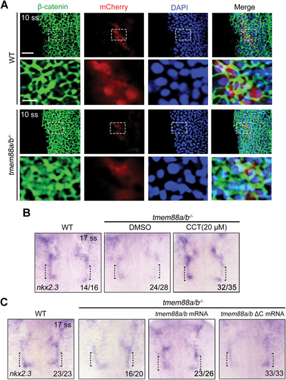
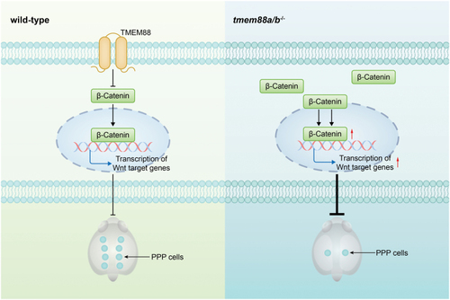
BIO treatment disturbs the morphogenesis of pharyngeal pouches. (A) Tg(TOPflash-GFP) embryos were treated with CCT036477 or BIO from bud stage to 24 hpf. Scale bar, 100 μm. CCT, CCT036477. (B) In situ hybridization analysis of nkx2.3 expression in embryos treated with DMSO or CCT036477 from the bud stage to 17 ss. (C) Heat shock-induced expression of Dkk1b in Tg(hsp70l:dkk1b-GFP) embryos. Heat shock was performed at the bud stage, and GFP fluorescence was imaged at 10 ss. Scale bar, 200 μm. (D, E) Tg(hsp70l:dkk1b-GFP) embryos were heat-shocked at the bud stage and harvested for in situ hybridization with the lef1 (D) or nkx2.3 (E) probe. (F) Comparative in situ hybridization analysis of nkx2.4b expression in wild-type embryos treated with or without BIO from the bud stage. (G, H) Wild-type embryos were exposed to different concentrations of BIO from the bud stage to 17 ss and harvested at 36 hpf for in situ hybridization with the nkx2.3 (G) or cv2 (H) probe. The ratios of affected embryos were indicated. (I) Tg(nkx2.3:mCherry) embryos were treated with DMSO or 10 μM BIO from the bud stage to 17 ss. Subsequently, these embryos were harvested at 36 hpf for in vivo confocal imaging. Scale bars, 100 μm.

Excessive Wnt signal compromises PPP specification. (A) Expression of nkx2.3 in embryos treated with DMSO or BIO. The black dotted lines indicate the region where the PPPs are located. (B, C) Representative confocal sections showing Tg(nkx2.3:mCherry) embryos treated with DMSO or BIO (B). The PPPs are indicated by white dotted lines. Scale bar, 20 μm. Quantitative analysis of PPP cell numbers is shown in (C). The group values are expressed as mean ± SD. (D, E) Confocal images illustrating the specification of nkx2.3+ pouch progenitors from the pharyngeal endoderm. White arrowheads indicate the presence of GFP+/mCherry+ cells in Tg(nkx2.3:mCherry;sox17:GFP) embryos (D). Scale bar, 20 μm. Ratio of mCherry+ PPPs to the GFP+ pharyngeal endoderm of embryos treated with DMSO or BIO was achieved from three independent assays (E). The group values are expressed as mean ± SD.

Tmem88 deletion severely impedes PPP specification. (A) In situ hybridization of tmem88a and tmem88b in wild-type embryos. (B, C) Double-color fluorescence in situ hybridization showing the expression of mCherry (red) and tmem88a or tmem88b (green) in Tg(nkx2.3:mCherry) embryos at the 10 (B) and 17 ss (C). The white arrow heads indicate the mCherry+ PPPs that do not express tmem88a or tmem88b. Scale bar, 50 μm. (D) Expression of nkx2.3 in wild-type and tmem88a/b−/− embryos at the 17 ss. The black dotted lines indicate the region where the pouch progenitors are located. (E, F) Representative confocal sections showing mCherry+ PPPs in wild-type and tmem88a/b−/− embryos at 17 ss (E). The PPPs are indicated by white dotted lines. Scale bar, 50 μm. Quantification of the number of pericardial and pouch progenitors positive for mCherry in wild-type and tmem88a/b−/− mutants is shown in (F). The group values are expressed as mean ± SD. (G, H) Requirement of Tmem88 for pouch progenitor specification. Wild-type and tmem88a/b−/− mutant embryos in the Tg(nkx2.3:mCherry;sox17:GFP) background were imaged at 10 ss (G). Scale bar, 20 μm. Ratio of mCherry+ PPPs to the GFP+ pharyngeal endoderm of wild-type or tmem88a/b−/− embryos was quantified from three independent assays (H). The group values are expressed as mean ± SD.

Tmem88 deletion has minor effect on pharyngeal pouch morphology. (A, B) The expression of nkx2.3 (A) and cv2 (B) in wild-type and tmem88a/b−/− mutants at 36 hpf. (C) Wild-type and tmem88a/b−/− mutant embryos in the Tg(sox17:GFP) background were imaged at 36 hpf. Scale bars, 20 μm. (D) Zn8-labeled pharyngeal pouches in both wild-type and tmem88a/b−/− mutants at 36 hpf. Scale bars, 50 μm.

Tmem88 promotes PPP specification through inhibiting Wnt/β-catenin signaling. (A) The subcellular localization of β-catenin in PPP cells of both wild-type and tmem88a/b−/− embryos in the Tg(nkx2.3:mCherry) background. Embryos were stained with the indicated antibodies. Nuclei were counterstained with DAPI (blue). The boxed area in the upper image (scale bar, 20 μm) is presented at a higher magnification in the corresponding lower image (scale bar, 100 μm). (B) Expression of nkx2.3 in wild-type or tmem88a/b−/− embryos treated with or without 20 μM CCT036477. (C) Expression of nkx2.3 in wild-type or mutant embryos injected with indicated mRNAs at 17 ss.

A schematic diagram showing the regulatory mechanism, whereby TMEM88 promotes PPP specification through suppressing Wnt/β-catenin signaling. The deficiency of tmem88a/b leads to an excessive accumulation of β-catenin in both the cytoplasm and nucleus of pharyngeal endodermal cells that are intended to differentiate into PPPs, thereby resulting in hyperactivation of Wnt signaling and defects in PPP specification.

Acknowledgments
This image is the copyrighted work of the attributed author or publisher, and ZFIN has permission only to display this image to its users. Additional permissions should be obtained from the applicable author or publisher of the image. Full text @ Life Med