|
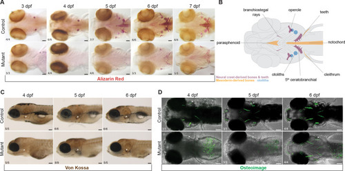
Mineralization deficit in zebrafish sox10 mutants. (A) Example images illustrating a major delay in initiation of bone mineralization in sox10 mutants between 3 and 7 dpf revealed by Alizarin Red staining. Some mineralization is present by 5 dpf but never achieves control levels before lethality at 8 dpf. (B) Schematic representation of the affected mineralized structures and their embryonic origins. (C,D) Von Kossa (C) and OsteoImage (D) staining showing absent calcium deposition and hydroxyapatite formation in sox10 mutants at 4 dpf and gradual recovery starting at 5 dpf. Arrowheads point to the opercle (op). Numbers in panels indicate the proportion of larvae of that genotype with the presented phenotype. Scale bars: 100 µm.
|