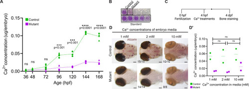
sox10 mutants have a severe whole-body calcium deficit. (A,B) Colorimetric calcium assay reveals significantly lower levels of Ca2+ in sox10 mutants after mineralization begins at 3 dpf. Each data point represents a pool of 10-15 embryos. Different shapes represent biological replicates assayed on different days (unpaired t-tests; 36 hpf: P=0.580, d.f.=8; 48 hpf: P=0.083, d.f.=8; 72 hpf: P=0.091, d.f.=7; 96 hpf: P=0.0002, d.f.=6; 120 hpf: P=0.0008, d.f.=4; 144 hpf: P=0.000008, d.f.=4; 168 hpf: P=0.000005, d.f.=6). B shows an example of the colorimetric assay, showing a clear reduction in mutants. (C) Schematic of the Ca2+ treatment protocol. (D,D′) Increasing ambient Ca2+ levels to 2 or 10 mM does not rescue the mineralization deficit (D) or Ca2+ content (D′) (unpaired t-tests; 1 versus 2 mM: P=0.963, d.f.=2; 1 versus 10 mM: P=0.778, d.f.=2; 2 versus 10 mM: P=0.748, d.f.=2). Numbers in panels indicate the proportion of larvae of that genotype with the presented phenotype. In D′, ratios reflect the number of imaged larvae of that genotype with the presented phenotype, and dashed lines indicate the median. Significance determined by unpaired t-test. ns, not significant. Scale bars: 100 µm.
|
