FIGURE

Fig 5

ID
ZDB-FIG-250318-15
Publication
Dicipulo et al., 2025 - Functional role for Taz during hindbrain ventricle morphogenesis
Other Figures
All Figure Page
Back to All Figure Page
Fig 5

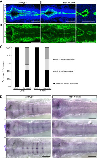
Apicobasal polarity components and patterned gene expression is perturbed in taz-/- mutants.

Changes to apicobasal polarity and cytoskeletal organization were assayed in wild-type and taz-/- mutants. (A) Crb2a localization in wild-type animals outlines the apical side of the ventricle. In taz-/- mutants, this localization is disturbed at locations where the ventricle fails to separate. Cell nuclei labelled with TO-PRO-3. (B) Phalloidin staining of F-actin in wild-type animals is seen strongly at the apical side of the ventricle lips, whereas in taz-/- mutants, locations where failure of midline separation occurs, we observe disturbances to F-actin organization. (C) Quantification of embryos observed having continuous, apposed, or gaps in apical localization. In wild-type animals, apical localization of Crb2a and F-actin is seen continuously from the anterior to posterior axis of the hindbrain ventricle, whereas in taz-/- mutants, a large proportion show either a gap or apposition of apical surfaces. (D) In taz-/- mutants, expression of genes that appear in a striated pattern are lost. These genes include rfng, rasgef1ba, and rac3b.

Expression Data

Expression Detail
Antibody Labeling
Phenotype Data

Phenotype Detail
Acknowledgments
This image is the copyrighted work of the attributed author or publisher, and ZFIN has permission only to display this image to its users. Additional permissions should be obtained from the applicable author or publisher of the image. Full text @ PLoS One