FIGURE SUMMARY
Title

Functional role for Taz during hindbrain ventricle morphogenesis

Authors
Dicipulo, R., Selland, L.G., Carpenter, R.G., Waskiewicz, A.J.
Source
Full text @ PLoS One

taz-/- mutants show midline separation defects.

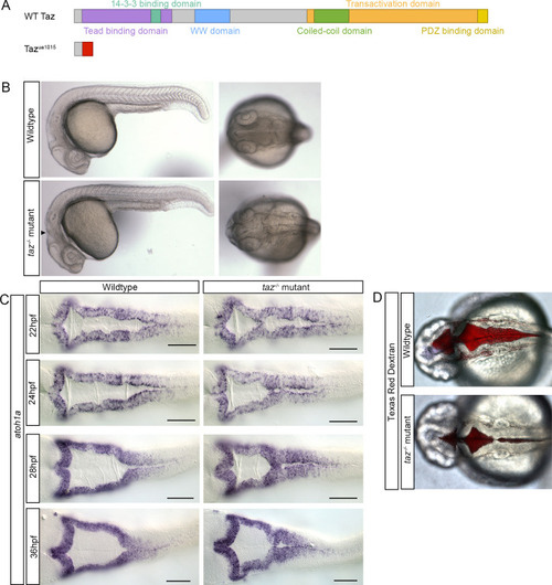
(A) Predicted transcripts of wild-type (top) and the TALEN generated
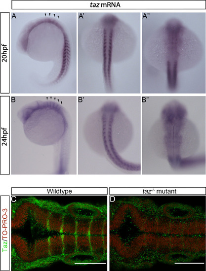
 tazua1015 mutant allele (bottom). The zebrafish tazua1015 mutant allele contains a 29 bp deletion, 25 bp downstream from the start codon, which results in a frameshift mutation and loss of all functional domains. (B) Microscopic analysis of embryonic phenotypes. Wild-type (top) and taz-/- mutant embryos (bottom) were analyzed at 24 hpf, shown at both lateral and dorsal views. Consistently, taz-/- mutants display disruptions to hindbrain morphology (lateral view, arrowhead) as well as failure of the brain ventricle midline separation (dorsal view, arrowhead). (C) atoh1a in situ hybridization on wild-type and taz-/- mutant embryos from 22-36 hpf were performed to visualize ventricle perturbations over the course of development. (D) Texas Red Dextran injections were performed into the ventricle of wild-type and taz-/- mutant embryos at 24 hpf to visualize extent of ventricle defects in live animals. Scale bar =  100 µm.

taz mRNA and Taz protein are seen in the hindbrain during BVS development.

(A-A”). In situ hybridization of wild-type embryos stage 20 hpf show taz expression throughout the embryo, from the trunk and tail to broadly in the rostral region, shown both laterally (A) and dorsally (A’, A”). (A’) Dorsal view of the embryo showing taz expression in the somite region. (A”) Dorsal view of the embryo hindbrain showing taz expression is enriched at rhombomere boundaries. (B-B”). At 24 hpf, broad expression of taz in the anterior and in the trunk is still seen; taz mRNA enrichment at rhombomere boundaries also persists. (C) Taz protein is localized to rhombomere boundaries in wild-type 24 hpf embryos whereas (D) taz-/- mutants display only background signal.

Wnt components are modified in taz-/- mutants and inhibition of

β-catenin mediated transcription results in reduced ventricle size. (A)
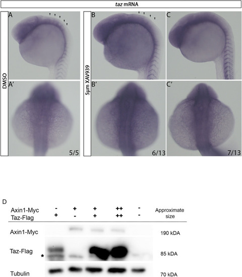
 in situ hybridization of wnt1 shows that in wild-type animals wnt1 expression is seen as striations along the hindbrain ventricle. In taz-/- mutants, this expression is disorganized. (B) Wild-type embryos were treated with 200 µM ICG-001 and 100 µM Windorphen to inhibit β-catenin mediated transcription. Compared to DMSO controls, treated embryos show reductions in ventricle size. (C) Similarly, inhibition of β-catenin mediated transcription via injection of 10 pg of a drTCF3 resulted in a reduced ventricle size compared to controls injected with eGFP alone. Cell nuclei labelled with TO-PRO-3. (D) Compared to wild-type animals, treatment with 5 µM XAV939 reduced ventricle size as visualized using atoh1a in situ (top). Additionally, XAV939 treated animals show a reduction of Taz protein localization at rhombomere boundaries (bottom). Cell nuclei labelled with TO-PRO-3. (E) Animals treated with 10 µM SB216763 show little change to ventricle shape and size compared to wild type animals, visualized using atoh1a in situ (top). However, SB216763 animals show an increase in Taz signal throughout the rhombomere, as seen more clearly in the black and white images of SB216763 treated animals included. Cell nuclei labelled with TO-PRO-3. (F) Treatment with 10 µM SB216763 results in 75% increase in fluorescence (P-value ≤  0.001) within rhombomeres, and an increase in fluorescence of 145% at rhombomere boundaries (P-value ≤  0.001). (G) Wild-type embryos were treated with 200 µM ICG-001 and 100 µM Windorphen to inhibit β-catenin mediated transcription. Compared to DMSO controls, treated embryos show reductions in ventricle size. Compared to DMSO controls, treated embryos show little to no reduction in Taz immunofluorescence. Scale bars =  100 µm.

Wnt activity affects taz expression at rhombomere boundaries.

(A-C’) In DMSO treated animals, at 24 hpf taz mRNA is seen broadly in the anterior of the embryo, with visible striations indicative of taz mRNA enrichment at rhombomere boundaries shown laterally (A) and dorsally with the hindbrain in focus (A’). In 5 µM XAV939 treated animals, at 24 hpf show a reduction of taz mRNA enrichment at the rhombomere boundaries (B-B’) or complete loss of this striated pattern (C-C’). (D) Injection of 200 pg Taz-Flag allowed visualization of the expected band for via western blot (lane 1; note the appearance of a non-specific band labeled *  present in all lanes, including the lane 5 negative control). Similarly, injection of 200pg Axin1-Myc resulted in a band at the expected MW being visualized via 9E10 western blot (lane 2). Co-injection of 200 pg Axin1-Myc and 200 pg Taz-Flag resulted in a greater amount of Flag-Taz detected (lane 3) which was also seen when 400 pg of Axin1-Myc and 400 pg Taz-Flag was injected (lane 4).

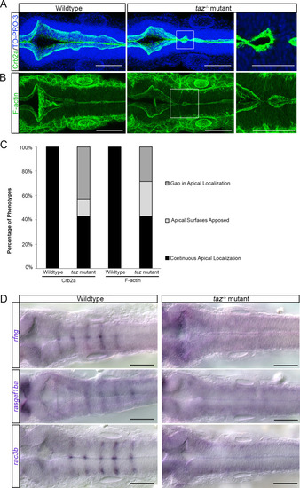
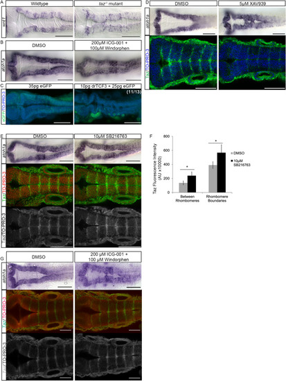
Apicobasal polarity components and patterned gene expression is perturbed in taz-/- mutants.

Changes to apicobasal polarity and cytoskeletal organization were assayed in wild-type and taz-/- mutants. (A) Crb2a localization in wild-type animals outlines the apical side of the ventricle. In taz-/- mutants, this localization is disturbed at locations where the ventricle fails to separate. Cell nuclei labelled with TO-PRO-3. (B) Phalloidin staining of F-actin in wild-type animals is seen strongly at the apical side of the ventricle lips, whereas in taz-/- mutants, locations where failure of midline separation occurs, we observe disturbances to F-actin organization. (C) Quantification of embryos observed having continuous, apposed, or gaps in apical localization. In wild-type animals, apical localization of Crb2a and F-actin is seen continuously from the anterior to posterior axis of the hindbrain ventricle, whereas in taz-/- mutants, a large proportion show either a gap or apposition of apical surfaces. (D) In taz-/- mutants, expression of genes that appear in a striated pattern are lost. These genes include rfng, rasgef1ba, and rac3b.

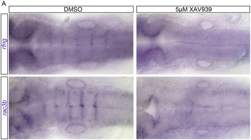
Wnt activity affects boundary cell gene expression.

In DMSO treated animals at 24 hpf rfng mRNA is expressed at rhombomere boundaries (15/15). In 5 μM XAV939 treated animals at 24 hpf this striated expression pattern is lost (23/23). In DMSO treated animals at 24hpf rac3b is also expressed at boundaries (16/16). In 5 μM XAV939 treated animals at 24 hpf this striated expression pattern is lost (23/23).

Acknowledgments
This image is the copyrighted work of the attributed author or publisher, and ZFIN has permission only to display this image to its users. Additional permissions should be obtained from the applicable author or publisher of the image. Full text @ PLoS One