
Fig. 8
- ID
- ZDB-FIG-250318-62
- Publication
- Zhou et al., 2025 - Effect of Dync1h1 on Phototransduction Protein Transport and the Development and Maintenance of Photoreceptor Cells in Zebrafish
- Other Figures
- All Figure Page
- Back to All Figure Page
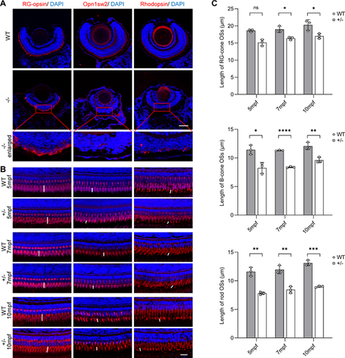
Transport disorder of phototransduction proteins in PRCs due to dync1h1-deficiency. (A, B) Immunofluorescence images of WT and dync1h1−/− retinas at 5 dpf, and WT and dync1h1+/− retinas at indicated ages, with anti-RG-opsin antibody, anti-Opn1sw2, and anti-Rhodopsin antibodies marking blue-cone opsin, red/green-cone opsin, and rhodopsin, respectively. Nuclei are stained by DAPI. Zoom-in pictures were shown in the red rectangle. The lengths of OS were labeled with white vertical lines. (C) Quantitative analysis of the lengths of RG-cone OS, B-cone OS, and rod OS in WT and dync1h1+/− retinas at indicated ages. +/−, dync1h1 heterozygote; −/−, dync1h1 homozygote. Scale bar = 50 µm (A), 20 µm (B). Data are shown as mean ± SD. Not significant (ns), P > 0.05; *, P < 0.05; **, P < 0.01; ***, P < 0.001; ****, P < 0.0001, n = 3 biologically independent samples per group. DAPI, 4′,6-diamidino-2-phenylin-dole; dpf, days post fertilization; H&E, hematoxylin and eosin; OS, outer segment; PRC, photoreceptor cell; WT, wild type; SD, standard deviation. |