
Fig. 10
- ID
- ZDB-FIG-250318-64
- Publication
- Zhou et al., 2025 - Effect of Dync1h1 on Phototransduction Protein Transport and the Development and Maintenance of Photoreceptor Cells in Zebrafish
- Other Figures
- All Figure Page
- Back to All Figure Page
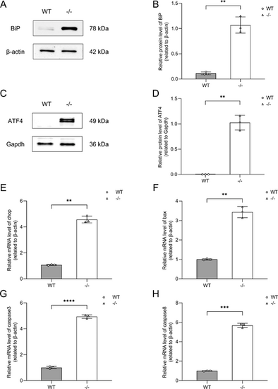
Validation of ER stress-induced apoptosis pathway in vivo. (A) Western blot analysis of BiP expression in WT and dync1h1−/− zebrafish at 5 dpf, with β-actin as the endogenous control. (B) Quantitative analysis showing elevated BiP levels in dync1h1−/− compared to WT. (C) Western blot analysis of ATF4 protein expression in WT and dync1h1−/− zebrafish at 5 dpf, with Gapdh as endogenous control. (D) Quantitative analysis showing elevated ATF4 protein levels in dync1h1−/− compared to WT. (E–H) The qRT-PCR analysis showing increased mRNA levels of chop, bax, caspase3, and caspase8 in dync1h1−/− relative to WT. β-actin was used as the endogenous control. −/−, dync1h1 homozygote. Data are shown as mean ± SD. **, P < 0.01; ***, P < 0.001; ****, P < 0.0001, n = 3 biologically independent samples per group. ATF 4, activating transcription factor 4; WT, wild type; BiP, binding immunoglobulin protein; ER, endoplasmic reticulum; qRT-PCR, quantitative real-time polymerase chain reaction; SD, standard deviation. |