
Fig. 2
- ID
- ZDB-FIG-250407-28
- Publication
- Li et al., 2024 - Dehydroervatamine as a promising novel TREM2 agonist, attenuates neuroinflammation
- Other Figures
- All Figure Page
- Back to All Figure Page
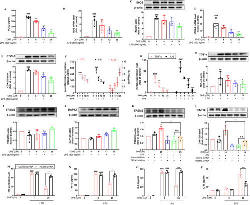
DHE suppressed LPS-induced activation of proinflammatory mediators and prevented the release of inflammatory cytokines by targeting TREM2. Cells were pretreated with DHE for 1 h and then exposed to LPS for another 24 h. (A) PGE2 production was determined by ELISA; n = 4. (B, D) The mRNA levels of iNOS and COX-2 were detected by RT-PCR analysis; n = 3. (C, E) Protein levels of iNOS and COX-2 were measured by Western blot analysis; n = 3. (F) Secretion levels of TNF-α, IL-6, and IL-10 in the cell culture medium were determined by relative ELISA; n = 4. (G) The mRNA levels of TNF-α and IL-6 were assessed by RT-PCR analysis; n = 3. (H) Protein levels of TNF-α were evaluated by Western blot analysis; n = 3. TREM2 (I) and DAP 12 (J) protein expression were detected by Western blot analysis and quantified by ImageJ. After TREM2 was knocked down using shRNA, microglial cells were treated with DHE in the presence or absence of LPS to determine TREM2 (K) and DAP12 (L) protein expression levels. The supernatant of BV2 cells was then used to measure NO production (M), TNF-α (N), IL-6 (O), and IL-10 (P) via Griess reagent and ELISA assay, respectively; n = 3–4. Data are presented as the mean ± SD analyzed by one-way ANOVA. ∗p < 0.05, ∗∗p < 0.01, and ∗∗∗p < 0.001 vs. the LPS-treated group; #p < 0.05, ##p < 0.01, and ###p < 0.001 vs. the control group; N.S. = not significant. |