|
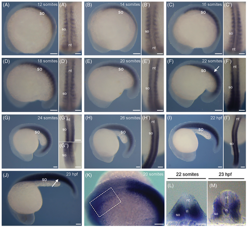
Spatio-temporal expression of rbm24b transcripts during zebrafish development. (A–K) Lateral views, with anterior to the left, of whole-mount embryos from 10-somite stage to 24 hpf, showing rbm24b expression in the developing somites. (A′–I′) Dorsal views, with anterior to the top, of embryos shown in A-I, focused on the developing somites. (K) Weak rbm24b hybridization signal (outlined by dashed rectangle) can be observed in the heart area with prolonged incubation period for chromogenic reaction. (L) Transverse section of the embryo at 20-somite stage shown in F, at the level of somite (white arrow). (M) Transverse section of the embryo at 23 hpf shown in J, at the level of the somite (white arrow). So, somite; nt, neural tube; n, notochord. Scale bars: (A–K) 100 μm; (A′—I′, L, M) 50 μm.
|