
FIGURE
Fig. 9
- ID
- ZDB-FIG-250527-9
- Publication
- Saquet et al., 2024 - Knockout of rbm24a and rbm24b genes in zebrafish impairs skeletal and cardiac muscle integrity and function during development
- Other Figures
- All Figure Page
- Back to All Figure Page
Fig. 9
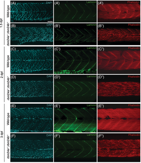
Formation of myotendinous junction in rbm24a and rbm24b mutants. (A–F) DAPI staining. (A′–F′) Laminin immunofluorescence staining. (A″–F″) Phalloidin staining. Lateral view for all embryos, with anterior to the right. Notice that myofibers appear dispersed and disorganized in rbm24a and rbm24b mutant embryos, whereas the myotendinous junction of somite boundaries exhibit a pattern similar to wild-type embryos. At each developmental stage, 15 to 20 embryos per genotype were analyzed. Scale bar: 25 μm. |
Expression Data
Expression Detail
Antibody Labeling
Phenotype Data
| Fish: | |
|---|---|
| Observed In: | |
| Stage Range: | Prim-25 to Protruding-mouth |
Phenotype Detail
Acknowledgments
This image is the copyrighted work of the attributed author or publisher, and
ZFIN has permission only to display this image to its users.
Additional permissions should be obtained from the applicable author or publisher of the image.
Full text @ Dev. Dyn.