
Fig. 5
- ID
- ZDB-FIG-250602-93
- Publication
- Liu et al., 2025 - Rhodopsin Induces Myopia via Lipid Peroxidation in Zebrafish Reared in a Dark Environment
- Other Figures
- All Figure Page
- Back to All Figure Page
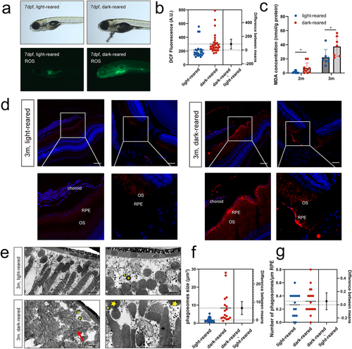
Lipid peroxidation in the retinas of zebrafish reared under a dark environment (a) Representative images of bright fields and fluorescence for ROS detection by DCFH-DA incubation. (b) Statistical analysis of DCF fluorescence by estimation plot; n = 23 for the light-reared and n = 33 for the dark-reared group. (c) MDA concentrations in zebrafish eyeballs in the two groups. n = 11 for the light-reared and n = 10 for the dark-reared group at 2 months; n = 7 for the light-reared and n = 7 for the dark-reared group at 3 months. (d) 4-HNE immunofluorescence of the retina and optic nerve. (e) Transmission electron microscopy of the outer segment of rod cells of 3-month-old adult fish. Yellow stars represent phagosomes, while red arrows refer to the discoid structure of the outer segment of the rod cells. (f, g) Estimation plot showing the number and size of phagosomes, respectively. Statistical significance was determined using the Student's t-test. The scale bars are 50 μm. 4-HNE, 4-hydroxynonenal; DCF, dichlorofluorescein; DCFH-DA, 2′,7′-dichlorodihydroxyoxalol diacetate; MDA, malondialdehyde. *p < 0.05. |