|
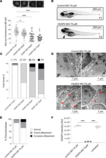
CKAP4 zebrafish homolog knockdown causes proteinuria and podocyte FPE. CKAP4 was knocked down in zebrafish using MO in different concentrations, 30, 50, and 75 μM MO. Proteinuria was measured by reduction of eye fluorescence (A). Example of normal phenotype (P1, no edema) and fish with mild edema (P2) in CKAP4 MO; the arrow points at pericardial edema (B). Assessment of fish phenotypes in the different groups; from P1 to P4 (severe edema) was conducted with reference to Hanke et al. (25, 27) and Ursu et al. (26) (C). Representative electron microscopy pictures of the filtration barrier of zebrafish glomeruli from control MO and CKAP4 MO (75 μM). Control MO shows normal podocyte foot processes (this pattern is indicated with short white arrows) while the CKAP4 MO shows podocyte FPE (partial effacement patterns are indicated with red arrows, complete effacement with short red arrows) (D). Quantification of podocyte FPE percentages in control MO and CKAP4 MO shows increased effacement in CKAP4 MO–treated zebrafish (E). Validation of CKAP4 MO knockdown of CKAP4 as obtained via proteomics (F). A: a minimum of 32 larvae per group was used. Error bars: average ± SEM. *P < 0.05, ***P < 0.001, Mann-Whitney test. E: n = 336 (control MO), n = 319 (CKAP4 MO) foot processes were counted, from n = 14 (control MO) and n = 8 (CKAP4 MO) independent images per group. F: 3 independent experiments were performed; each treatment in each replicate is derived from lysate of a minimum of 8 pooled embryos. Error bars in both panels represent average ± SEM. ***P < 0.001, 1-way ANOVA with Tukey’s multiple-comparison test. CKAP4, cytoskeleton associated protein 4; MO, morpholino.
|