|
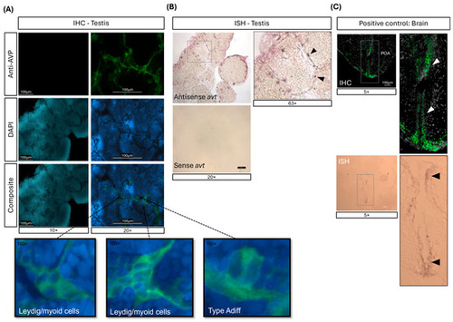
Localization of arginine vasotocin (AVT) expression in adult zebrafish testis detected by (A) immunohistochemistry (IHC) and (B) in situ hybridization (ISH). (A) IHC on testis sections showing AVT-positive staining (top), DAPI nuclear staining (middle) and composite images (bottom). The left panel shows a negative control (no primary antibody), while the right panel shows specific AVT labeling. Inserts highlight cellular localization. (B) ISH on testis sections incubated with antisense (top) and sense (bottom) avt RNA probes. Cellular localization of avt is indicated by arrowheads. (C) IHC (top) and ISH (bottom) of the forebrain, used as positive control. The arrowheads indicate AVT-positive sites within the preoptic area (POA) of the hypothalamus.
|