
FIGURE 4
- ID
- ZDB-FIG-260618-83
- Publication
- Jansen et al., 2026 - Hnrnpa1 is essential for early zebrafish development and lipid metabolism: insights from a novel zebrafish knockout model
- Other Figures
- All Figure Page
- Back to All Figure Page
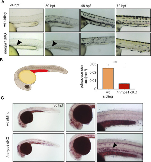
Hnrnpa1 dKOs have lipid metabolism defects. (A)hnrnpa1 dKO embryos have a thinned yolk extension. Images of wildtype (upper panel) and hnrnpa1 dKO (lower panel) embryos at 24 hpf, 30 hpf, and 48 hpf. At 24 hpf hnrnpa1 dKO can be hardly distinguished from their wildtype siblings apart from a small bulge in the yolk extension (arrowhead). After 30 hpf the yolk extension of hnrnpa1 dKO embryos starts to become thin, which becomes more severe over time resulting in a severely reduced yolk extension after 48 hpf up to 72 hpf. Lateral view. Anterior to the left. Images were taken with Axio Scope A1. (B) Schematic illustration of the measured area of the yolk extension (indicated in red) at 30 hpf which was used for quantification. Quantification of the two-dimensional yolk extension area at 30 hpf shows significant reduction in yolk extension area in hnrnpa1 dKO compared to wildtype. Student’s t-test (n = 13; ***p < 0.0001). Error bars indicate S.E.M. (C) Oil Red O stained the head structures, the yolk, yolk extension and structures around the spinal cord. Reduced amounts of neutral lipids in the yolk extension are present in hnrnpa1 dKO embryos at 30 hpf. Additionally, hnrnpa1 dKO embryos show increased uptake of neutral lipids to the trunk (arrowhead). Lateral view. Anterior to the left. Images were taken with Axio Scope A1 microscope. |
| Fish: | |
|---|---|
| Observed In: | |
| Stage: | Prim-15 |