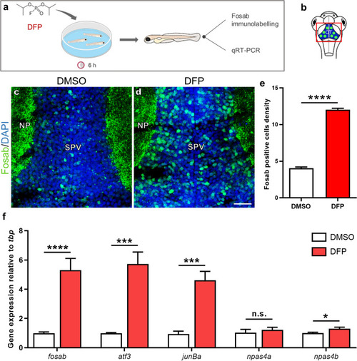
DFP exposure induces overexpression of the IEGs fosab, atf3, junBa, and npas4b. (a) As experimental set-up, 5 dpf larvae were exposed for 6 h to either 15 µM DFP or vehicle (1% DMSO) before processing for either Fosab immunostaining or brain dissection followed by RNA extraction and qRT-PCR analysis. (b) Scheme of a 5 dpf larva head with the red box showing the region of interest in the brain, uncovering the optic tectum (OT). (c,d) Fosab immunolabeling of optic tectum neurons in 5 dpf larvae exposed to either vehicle (c) or 15 µM DFP (d). Scale bar: 20 µm. (e) Quantification of Fosab-expressing neuron density in the optic tectum of 5 dpf larvae exposed to either vehicle (N = 3; n = 8) or 15 µM DFP (N = 3; n = 11) (unpaired t-test: ****, p < 0.0001). (f) qRT-PCR analysis of the accumulation of fosab, atf3, junBa, npas4a and npas4b RNAs relative to that of tbp in 5 dpf larvae exposed to either vehicle (n = 6) or 15 µM DFP (n = 6) (Student’s unpaired t-test: n.s., non-significant; *, p < 0.05; ***, p < 0.001; ****, p < 0.0001). N = number of larvae and n = number of slices. Abbreviations: NP, neuropil; SPV, stratum periventriculare.
|
