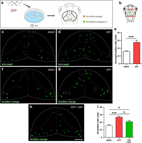
DFP exposure increased cell apoptosis. (a) As experimental set-up, 5 dpf larvae were exposed to either 15 µM DFP or vehicle (1% DMSO) for 6 h, before processing for either acridine orange (AO) staining or anti-activated caspase-3 (Act-casp3) immunolabeling. (b) Scheme of a 5 dpf larva head with the red box showing the region of interest in the brain, uncovering the optic tectum (OT). (c,d) Act-casp3 immunolabeling of OT neurons in 5 dpf larvae exposed for 6 h to either vehicle (c) or 15 µM DFP (d). (e) Quantification of Act-casp3-positive neurons in 5 dpf larvae exposed for 6 h to either 15 µM DFP (n = 12) or vehicle (n = 12) (Student’s unpaired t-test with Welch’s correction: ***, p < 0.001). (f–h) Visualization of AO-labeled apoptotic neurons in 5 dpf larvae exposed for 6 h to either vehicle (f), or 15 µM DFP (g) or 15 µM DFP + 40 µM diazepam (h). (i) Quantification of the number of acridine orange positive cells in 5 dpf larvae exposed for 6 h to either vehicle (n = 24), or 15 µM DFP (n = 17) or 15 µM DFP + 40 µM diazepam (DZP) (n = 10) (one-way ANOVA with Tukey’s multiple comparisons test: *, p < 0.05; ***, p < 0.001). Scale bar: 50 µm.
|
