
FIGURE 1
- ID
- ZDB-FIG-250407-43
- Publication
- Tatzl et al., 2025 - Deficiency of the Synaptic Adhesion Protein Leucine-Rich Repeat Transmembrane Protein 4 Like 1 Affects Anxiety and Aggression in Zebrafish
- Other Figures
- All Figure Page
- Back to All Figure Page
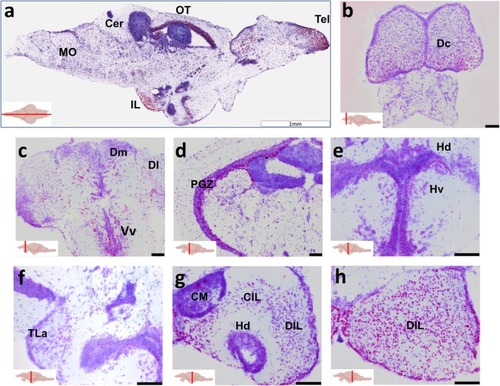
Lrrtm4l1 expression in the adult zebrafish brain. (a) Midsagittal section showing intense lrrtm4l1 expression (purple) in the telencephalon (Tel), inferior lobe (IL) and optic tectum (OT). (b–e) Coronal sections of the telencephalon (b, c), optic tectum (d), hypothalamus (e, f) and inferior lobe (g, h). Sections have been counterstained with thionine acetate for anatomical orientation. Scale bars are 1 mm in (a) and 100 μm in (b–h). The red line in the brain inserts indicates the sectioning plane. Cer, Cerebellum; CIL, central nucleus of the inferior lobe; CM, mamillary bodies; Dc, central zone of the dorsal telencephalon; DIL, diffuse nucleus of the inferior lobe; Dl, lateral zone of the dorsal telencephalon; Dm, Medial zone of the dorsal telencephalon; Hd, dorsal zone of the periventricular hypothalamus; Hv, ventral zone of the periventricular hypothalamus; IL, inferior lobe, MO, medulla oblongata; OT, optic tectum; PGZ, periventricular gray zone of optic tectum; Tel, telencephalon; TLa, torus lateralis; Vv, ventral zone of the ventral telencephalon. Schematic zebrafish brain inserts created by |