
Fig. 1
- ID
- ZDB-FIG-250729-88
- Publication
- Wu et al., 2025 - Mitochondrial dysfunction is driven by imbalanced fission and fusion of mitochondria in myofibrillar myopathy type 5
- Other Figures
- All Figure Page
- Back to All Figure Page
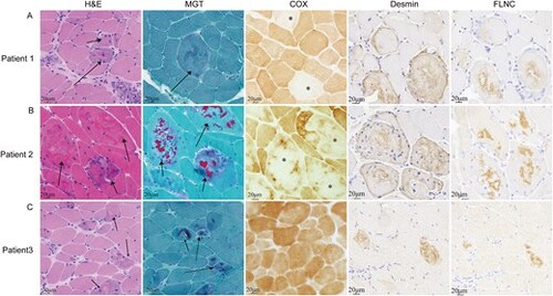
(A) Pathological biopsy and immunohistochemical staining results for patient 1. HE staining of patient 1 revealed mild variation in muscle fibre size, with basophilic material (indicated by arrows). Spherical bodies were observed via MGT staining (indicated by arrows), alongside COX-negative muscle fibres (indicated by *). (B) Pathological biopsy and immunohistochemical staining results for patient 2. Acidophilic material was evident on HE staining (indicated by arrows), cytoplasmic bodies on MGT staining (indicated by arrows), and large irregular areas of COX activity loss were noted within muscle fibres. (C) Pathological biopsy and immunohistochemical staining results for patient 3. HE staining revealed acidophilic material (indicated by arrows), cytoplasmic bodies on MGT staining (indicated by arrows), and no COX enzyme deficiency. Immunohistochemical staining revealed the deposition of desmin and filamin-C in all three patients. |