
Fig. 1
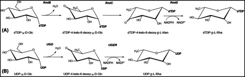
The L‐rhamnose biosynthetic pathway in prokaryotes and eukaryotes. (A) In Bacteria and Archaea, the biosynthesis of TDP‐β‐L‐rhamnose begins with the enzyme RmlB, which catalyzes the dehydration of TDP‐α‐D‐glucose, resulting in the formation of an unstable intermediate, TDP‐4‐keto‐6‐deoxy‐α‐D‐glucose. This intermediate then undergoes two subsequent enzymatic steps: a 3,5‐epimerization catalyzed by RmlC, and a stereospecific NADPH‐dependent 4‐keto‐reduction catalyzed by RmlD, ultimately yielding TDP‐β‐L‐rhamnose. Notably, Nematoda utilize a similar pathway, also employing the TDP‐bound substrate. (B) In contrast to the bacterial and archaeal pathway, most eukaryotes and giant viruses employ a slightly different strategy for L‐rhamnose biosynthesis. Here, the initial dehydration reaction, catalyzed by UGD (UDP‐D‐glucose 4,6‐dehydratase, a homolog of RmlB), utilizes UDP‐α‐D‐glucose as the substrate. The subsequent epimerization and reduction steps are typically carried out by a single, bifunctional enzyme resembling RmlD, termed UGER (UDP‐4‐keto‐6‐deoxy‐D‐glucose epimerase/reductase). Interestingly, higher plants exhibit further evolutionary adaptation, where the RmlB‐like and RmlD‐like domains are fused into single polypeptide chains known as RHM proteins. |