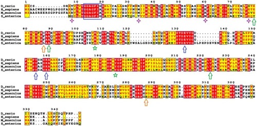
Sequence alignment of zebrafish Tgds with the corresponding orthologs from S. enterica and the human TGDS. Red boxes highlight the identical residues, while the yellow boxes refer to conserved ones. The blue box indicates the conserved Gly‐X‐X‐Gly‐X‐X‐Gly/Ala motif characteristic of a Rossmann fold. Residues forming the catalytic triad are marked with blue arrows, while green arrows point to conserved amino acids mutated in CMS and analyzed in this study. Orange arrows indicate the other mutations observed in CMS patients not analyzed in the present study. The green stars and purple diamonds denote the amino acids that establish contacts with the mutated residues and are discussed further in the Results section (‘Properties of the WT and mutant Tgds proteins’). The sequences shown are: S. enterica (UniProt Q9EU31, PDB 1G1A), H. sapiens (UniProt O95455) and D. rerio (GenBank NP_001428622). Protein sequence alignments were performed with Espresso on the T‐coffee server and figures were generated via ESPript.
|
