|
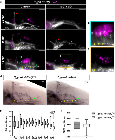
pax8 expression is downregulated in MCT8MO zebrafish embryos but is not involved in hindbrain vascular development. a Lateral view of maximum projections of the hindbrain vasculature structures at 32, 36 and 48 hpf in Tg(fli1:EGFP) in CTRMO and MCT8MO zebrafish embryos after WISH for pax8 (magenta) and immunostaining for GFP (endothelial marker, white). In CTRMO zebrafish embryos, pax8 expression appears in the posterior hindbrain region, juxtaposed to CtAs 4 to 7. In MCT8MO zebrafish embryos, pax8 expression was clearly reduced and only appears at 48 hpf (n = 9 – 18). Magnification of the selected area at 48 hpf for b CTRMO and c MCT8MO embryos are shown. d Lateral view of the hindbrain of Tg(pax8:dsRed)+/- (control group (pax8+/-)) and Tg(pax8:dsRed)-/- (hypomorph group (pax8-/-)) zebrafish embryos after WISH for cadherin-5 at 48 hpf. All 7 CtAs were present in control pax8+/- and hypomorph pax8-/- zebrafish embryos. Image J software was used to measure the length of e each CtA and f the PHBC between control pax8+/- (n = 18) and hypomorph pax8-/- (n = 14) zebrafish embryos. No changes were observed; Unpaired t-test (Mann-Whitney test). Data are presented as box-and-whisker plot, where the black thick horizontal line represents the median. The first and third quartiles are marked by the lower and upper edges of the boxes, respectively. Error bars represent standard deviations (smallest and highest value). For detailed statistics, see Supplementary Data 2. The red arrowhead indicates the mid-cerebral vein (MCeV), and the green arrowhead indicates the primordial hindbrain channels (PHBC). Yellow numbers 1 – 7 indicate the developed CtA in its respective rhombomere. Scale bar 50 μm for figures (a) and (d) and 20 μm for (b) and (c).
|