|
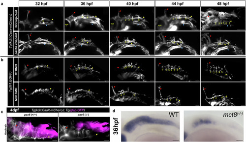
pax6a mutant zebrafish embryos show similar hindbrain vascular development defects as MCT8MO zebrafish embryos. a Live imaging of 32 hpf zebrafish embryo at the start of imaging between non-injected (control) and pax6a crispant zebrafish embryos are represented. The vascular system (kdrl) is shown in white (mCherry) in the reporter line Tg(kdrl:CaaX-mCherry). Dorsal view of maximum projection images of the mentioned time points is shown. b Live imaging of 32 hpf zebrafish embryo at the start of imaging between CTRMO and MCT8MO zebrafish embryos are represented. The vascular system is shown in white (GFP) in the reporter line Tg(fli1:EGFP). Dorsal view of maximum projection images of the mentioned time point is shown. n = 2. Scale bar: 50 μm. c Comparison between control (not injected) and mutant pax6a CRISPR zebrafish larvae at 4 dpf are presented. n = 5 (control), 9 (pax6a CRISPR). Mutant pax6a knockout zebrafish larvae present only 4 CtAs, while the control zebrafish have 7 CtAs. The red arrowhead represents the mid-cerebral vein (MCeV), the yellow arrowhead represents the primordial hindbrain channels (PHBC), and the blue arrowhead represents the lateral dorsal aorta (LDA). White * represents sprouting projections of the PHBC to the BA (due to the inclination of the hindbrain imaging, we can visualize these structures). Numbers 1 – 7 indicate the CtA in its respective rhombomere. d In mct8(-/-) embryos at 36 hpf it is observed a loss of pax6a hindbrain cells coincident with CtAs underdevelopment. Scale bar 50 μm.
|