|
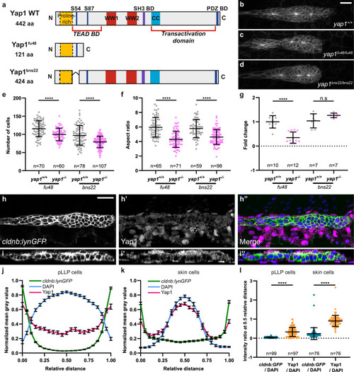
Tead-binding is essential for Yap1-mediated proliferation in the pLLP. a Schematic representation of the WT Yap1 and the mutant Yap1fu48 and Yap1bns22 proteins. Maximum intensity projection (MIP) of spinning disc confocal Z-stacks showing the pLLP in WT (b), yap1fu48 (c), and yap1bns22 (d) homozygous mutant embryos. Quantification of cell number (e) and aspect ratio (f) of the pLLP across genotypes (N = 3 independent replicates). g Relative yap1 transcript levels measured by qPCR in yap1+/+ and yap1−/− siblings carrying the fu48 or bns22 alleles at 32 hpf (N = 2 independent replicates). Single Z plan of confocal Z-stacks (h–h”) and orthogonal views (i–i”) showing Yap1 localization in 32 hpf WT embryos. Plot profile intensity across pLLP cells (j) and neighboring skin cells (k) in cldnb:lynGFP embryos at 32 hpf. Normalized mean gray values are shown for cldnb:lynGFP (membranes, green), DAPI (nuclei, blue), and Yap1 (magenta). l Quantification of normalized Yap1 and cldnb:lynGFP intensities relative to DAPI at 0.5 relative distance (N = 1 independent replicate). Unless otherwise indicated, the pLLP is marked by cldnb:lynGFP in all panels (b–d, h–i”). Data are presented as mean ± SD, except for (j, k), data are mean ± SEM. Unpaired t-tests (Mann–Whitney) were conducted. In all figures MIP are maximum intensity projections of spinning disc confocal Z-stacks. Scale bars: 20 μm (b–d, h–i”).
|